Deep phenotyping mycosis fungoides: profiling an orphan malignancy
As part of an ongoing collaboration with the Dermatology department of the Leiden University Medical Center (LUMC), CHDR’s Dermatology group has built an in-depth profile of mycosis fungoides, the most common form of cutaneous T-cell lymphoma. The project, which made use of CHDR’s DermaToolbox and the suction blister technique, also aimed to find predictive biomarkers for the therapeutic effect of chlormethine gel in this rare malignant skin condition.
‘The LUMC Dermatology department has a strong record of research into mycosis fungoides (MF) and keeps a central registry of patients with the disease,’ says Selinde Wind, Clinical Scientist at both CHDR and the LUMC. ‘MF patients from all over the Netherlands come to Leiden for diagnostics and treatment. In fact, the LUMC is one of the leading centres of expertise worldwide.’
Despite the impressive body of research, much is still unknown about this disease, which appears to be very heterogeneous on both a clinical and a molecular level. For example, it is not yet possible to predict whether a treatment will work in a particular patient. Wind: ‘In this project, which was supported by CHDR’s R&D Fund and co-funded by Recordati Rare Diseases and Helsinn, we therefore aimed to better phenotype MF. For this, we used the DermaToolbox, CHDR’s comprehensive dermatological testing suite.’
[text continues below the photo]
Initial findings
Preparations for this study already began back in 2020. Although MF is a rare diagnosis made only a few dozen times a year in the Netherlands, Wind was able to enrol 21 patients within five months, along with 10 matched healthy individuals. They were studied for about 6 months, which was extended to 12 months in a follow-up study. Wind: ‘I was working as a doctor in the outpatient clinic for MF patients at LUMC, so I could approach patients directly. This enabled us to include the required number of people in a very short time. In fact, it was a world record for a monocentre study of an orphan disease!’
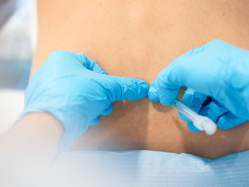
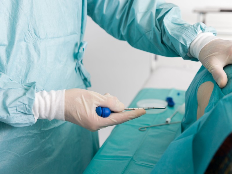
The team used a range of techniques to profile both the MF lesions and healthy skin of patients, as well as the skin of the matched healthy volunteers. This included imaging the blood flow and microvasculature and measuring the barrier function of the skin. ‘One technique we’ve used extensively here at CHDR is the suction blister. By drawing a blister locally on a lesion or on a healthy piece of skin and then sampling the interstitial fluid, you can get a good impression of the cells and proteins in the various layers of the skin,’ says Wind. ‘In MF, there is strong evidence that the tumour microenvironment is very important for the development of the disease. The suction blister technique allowed us to map that environment in detail.’
In addition to the ‘deep phenotyping’ of MF lesions, the DermaToolbox was also used to map the effect of treatment with chlormethine gel, a topical alkylating agent. Wind: ‘We wanted to investigate whether it’s possible to predict which patients will respond to treatment with chlormethine gel. This treatment can be effective in many patients, but there are also non-responders.’
Pioneering research
Currently, Wind is processing all the results of the project. Her first impressions are positive: ‘The fluid collected from the suction blisters has turned out to be a rich source of different types of cells. Besides tumour cells, we also found regulatory and cytotoxic T cells,’ says Wind. ‘The full analysis is still pending, but so far it seems that those different cell types may also be predictive of the treatment outcome. Maybe in the future we could even use the suction blister in the clinic, to select patients for this treatment.’
Interested in learning more?
More articles from Developing methods